Shoppers in labs and research centres are turning to laser capture microdissection for cleaner, targeted cell isolation; this roundup explains why the market is expanding, which systems and consumables matter, and how to pick the right setup for genomics, oncology or multi‑omics work.
Essential Takeaways
- Market growth: Laser capture microdissection is expanding rapidly, with steady revenue growth driven by spatial biology and personalised medicine needs.
- System types: Ultraviolet and infrared LCM, plus immunofluorescence‑guided and pressure‑catapulting systems, each offer trade‑offs in gentleness and precision.
- Consumables matter: Capture films and reagents are recurring costs but crucial for reproducible results; infrared systems can better preserve DNA/proteins.
- AI and software: Automation and AI image‑analysis are increasingly bundled to speed target selection and reduce manual bias.
- End users: Academic labs, hospitals, pharma and forensics are the primary buyers , choose for throughput and downstream workflows, not just headline specs.
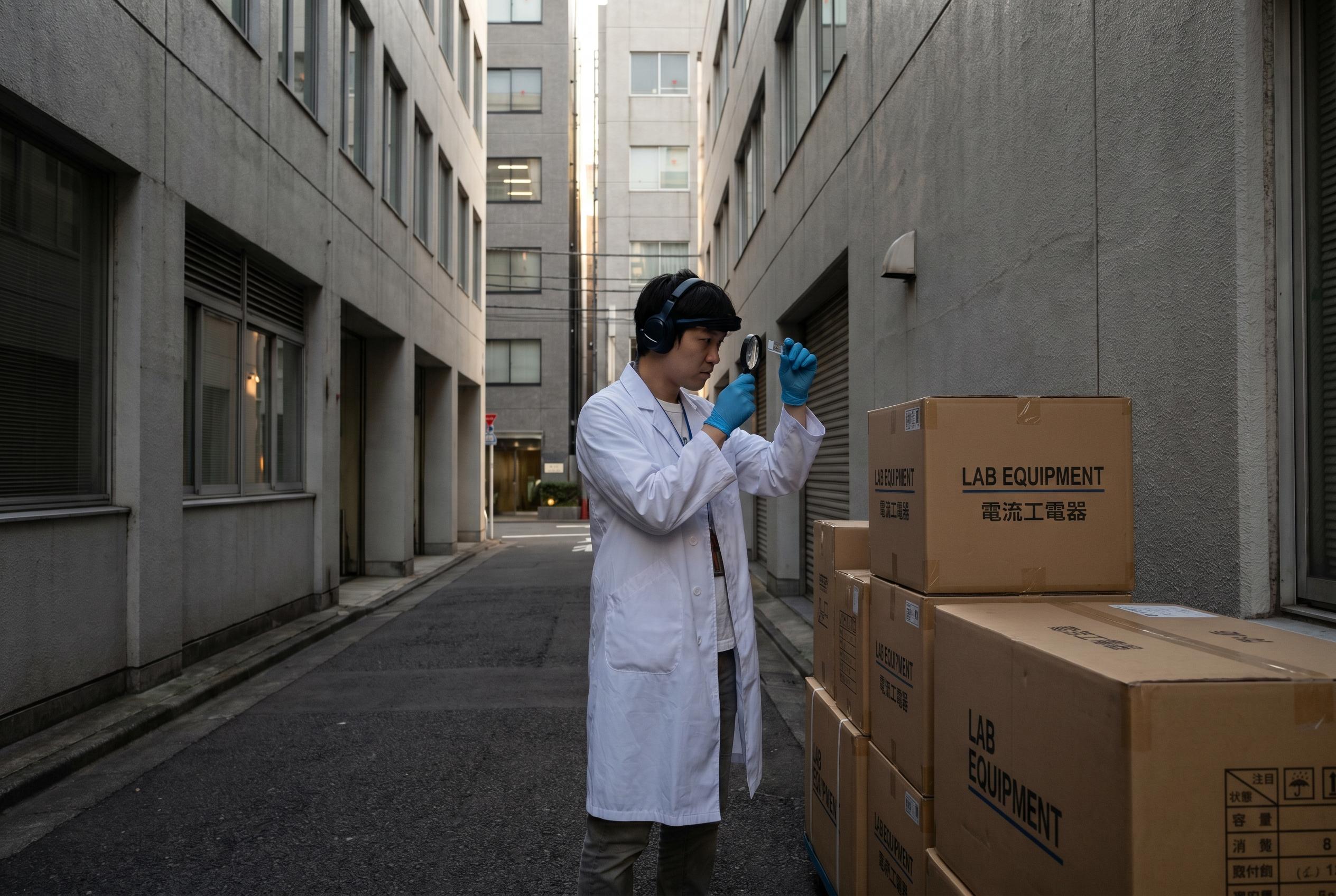
Why laser capture microdissection is suddenly everywhere
Spatial biology has put a spotlight on context: knowing which cell sat next to which matters as much as the genome inside it. Researchers told industry outlets that being able to pluck individual cells from tissue and keep their molecular cargo intact is transforming genomics and proteomics workflows. The result is practical , clearer signal in biomarker studies and fewer mixed‑cell artefacts in single‑cell analyses , and emotional: scientists feel they’re finally seeing tissues in the way biology intended.
Grand View Research and other market analysts note this shift is a major driver for market growth, as labs adopt LCM to support multi‑omics projects and precision oncology.
Picking a system: ultraviolet versus infrared (and the in‑between)
Not all LCM systems are created equal. Ultraviolet systems cut with high spatial precision, which is great for tiny targets, but they can be harsher on nucleic acids. Infrared systems use gentler capture films and are often preferred when DNA or proteins must remain intact. Immunofluorescence‑guided LCM helps if you’re hunting cells defined by markers rather than shape, while laser microdissection pressure‑catapulting is handy when you need sterile, contact‑free transfer.
Think about what you’ll analyse downstream. For sequencing, lean towards systems that preserve nucleic acid integrity. For proteomics or histology‑anchored work, prioritise gentle handling and compatibility with your staining protocols.
Consumables and software: the ongoing costs and the productivity lift
Consumables , capture films, special slides, reagents , are the unseen subscription that keeps LCM labs running. Analysts flag these items as reliable revenue for vendors and real budget items for labs. Plan yearly consumable budgets around your planned throughput, and factor in validation runs for new sample types.
Meanwhile, software is closing the usability gap. AI‑assisted workflows that pre‑identify target cells and suggest cut paths can shave hours off prep time and reduce operator variability. If you’re scaling up, invest in vendor software or third‑party image‑analysis packages that integrate with your LIMS.
Who’s buying and why it matters for your choice
The buyer mix is broad: universities and government institutes deploy LCM for basic science, hospitals use it for diagnostic research, and pharma and CROs adopt it for biomarker discovery and drug development. For clinical or regulated settings, certification, service contracts and traceability are as important as optical specs. For discovery labs, flexibility and compatibility with multiple downstream assays will be the deciding factors.
If you’re in a core facility, choose a mid‑range system with broad chemistry support; if you’re a focused oncology lab, a specialist immunofluorescence‑guided platform may deliver more immediate value.
Brands, competition and the innovation curve
The market features a mix of established instrument makers and specialist vendors. Big names are enhancing precision and usability, while niche companies push innovations in gentler capture methods and integration with spatial omics pipelines. Market reports show competition is healthy, which keeps prices competitive and accelerates new feature roll‑outs like AI guidance and improved capture consumables.
For buyers, that competition means you can negotiate service and consumable packages, and you aren’t locked into a single approach as your research evolves.
Choosing what’s right for your lab , a quick checklist
- Define your downstream assays first: sequencing, proteomics, histology.
- Match system type to sample sensitivity: infrared for gentleness, UV for micrometre precision.
- Budget for consumables and software maintenance, not just the instrument cost.
- Check vendor service, training and regulatory support if you’re in a clinical setting.
- Trial with your actual samples where possible; validation beats brochure specs every time.
It's a small change that can make every dissection safer and every downstream result clearer.
Source Reference Map
Story idea inspired by: [1]
Sources by paragraph: